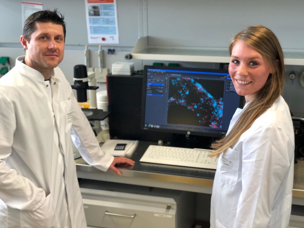
“Ilker, could you please pass the staining reagent to me? I will stain the breast cancer cells and then we both can have a look on it at the microscope!”
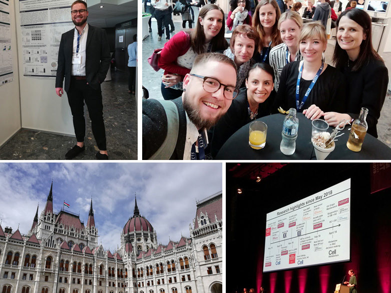
Lila Xu and Ilker A. Deniz have another very busy day in the lab. They have just started their Ph.D. thesis and are now part of an interdisciplinary team formed by the Tissue Engineering group (PI: Dr. Denis Corbeil) at the BIOTEC and the Department of Gynecology and Obstetrics (PIs: Prof. Pauline Wimberger and Dr. Jan Kuhlmann) of TU Dresden. For their thesis, both have one goal, they want to begin a fight against an old enemy for women – BREAST CANCER – and want to address important questions with their thesis:
Why can breast cancer be such a dangerous disease? Why do breast cancer cells migrate and form metastasis? How can breast cancer cells invade the bone and form bone metastasis? Do cancer cells have their own “feet” to walk?
To find answers, Lila and Ilker will look closer on breast cancer cells with different microscopy techniques, which enable observation down to tiny membrane structures. According to research results from the last years, Lila and Ilker know that breast cancer cells can shape their membranes to form their own „feet“, which helps them to leave the primary tumor site and to form metastasis. Scientists call them „ lamellipodia “ or „ magnupodia “. Both students will analyze these structures and investigate how breast cancer cells use their feet to invade the bone to form dangerous metastases.