By Ilker A. Deniza, Lila Bemmerleinb, Jana Karbanováa, Denis Corbeila, Jan D. Kuhlmannb, and Pauline Wimbergerb at aTissue Engineering Laboratories, Biotechnology Center (BIOTEC) and bDepartment of Gynecology and Obstetrics, Medical Faculty and University Hospital Carl Gustav Carus, Technische Universität Dresden, Dresden, Germany
The changes imposed on our society by the COVID-19 pandemic have affected everyone in a significant way. In Germany, as in most European countries, almost all research has been suspended (or reorganized as a home-office work), except for scientists and clinicians trying to combat and cure this highly contagious disease. While the curve is slowly flattening, we will soon be able to resume our work on cancer research.
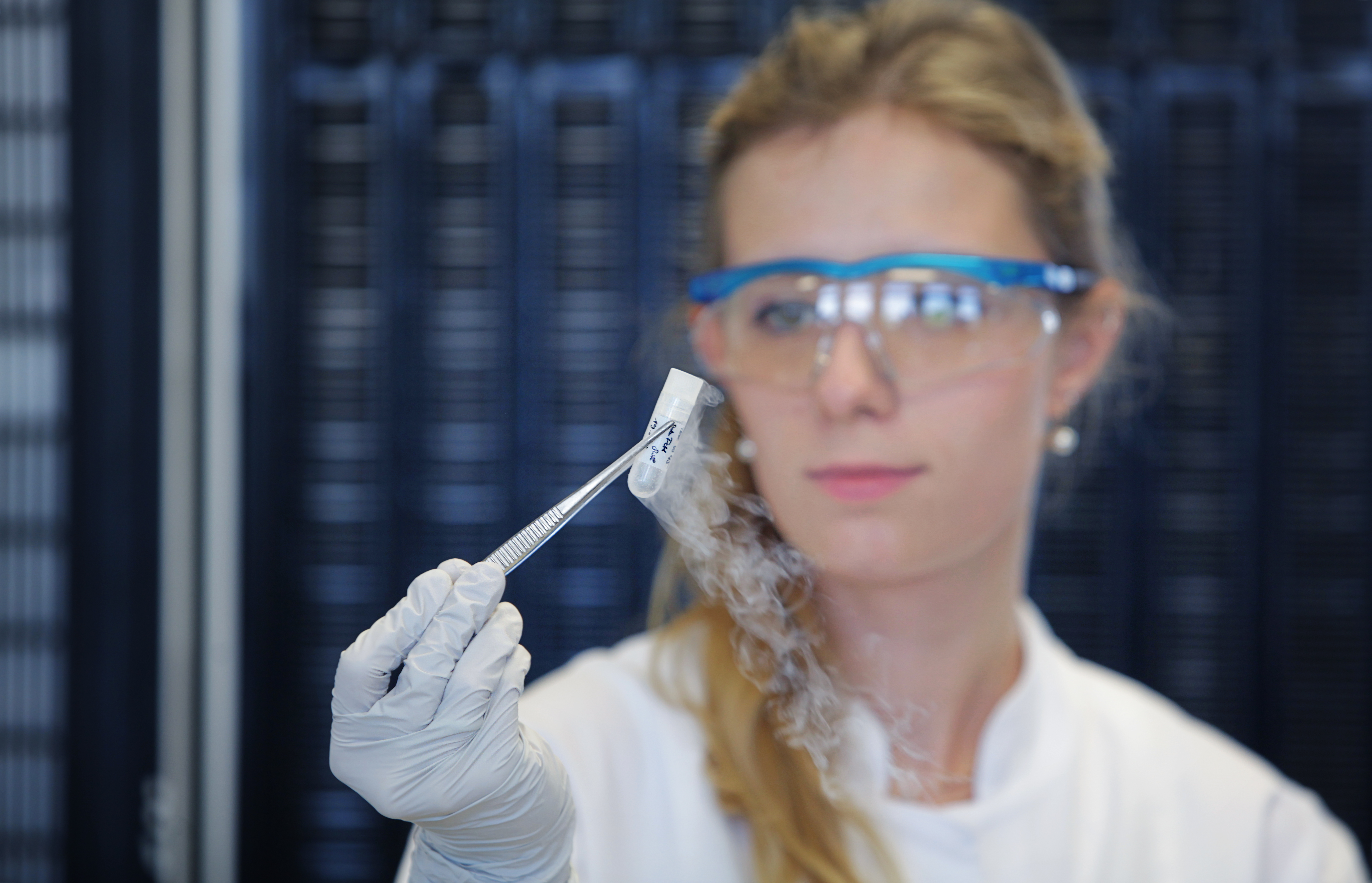
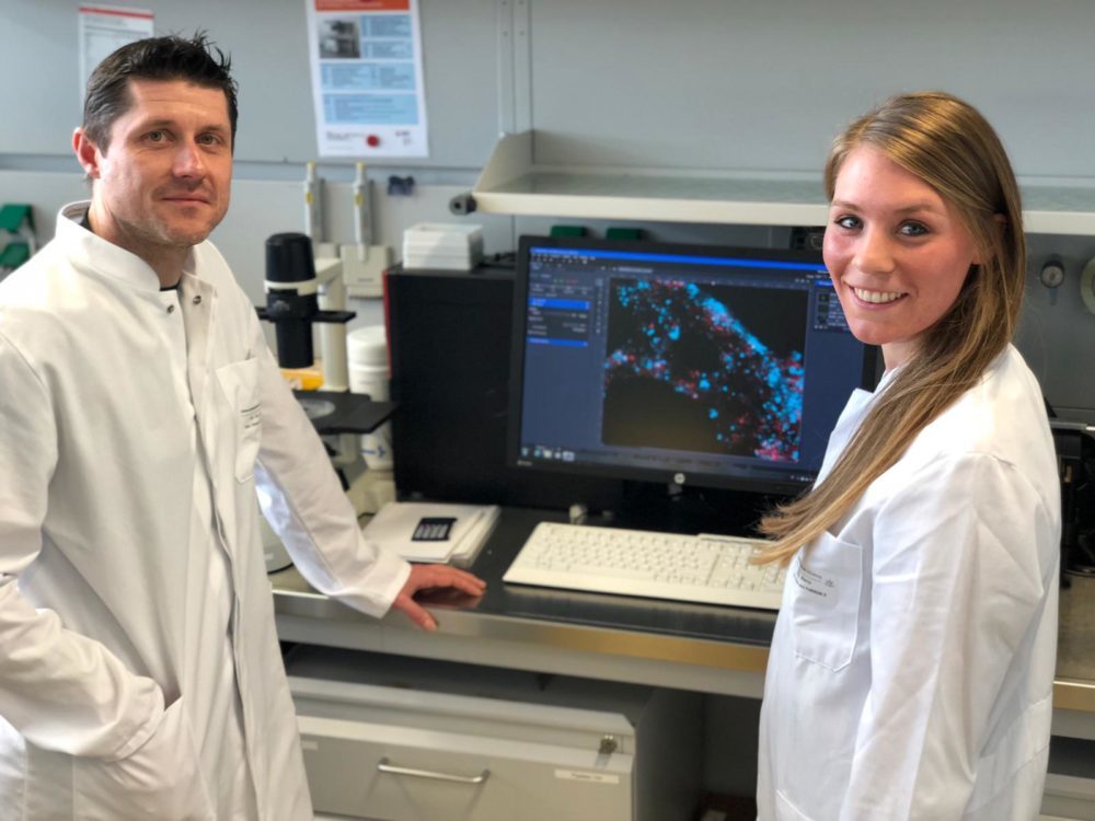
In our research team, we seek to understand how malignant cells disseminate to different organs – a process known as metastasis. What makes cancer such a life-threatening disease, is its unpredictability and its ability to adapt and thrive in seemingly unrelated parts of the body. To decipher how these cells can spread throughout the body, we use several microscopy techniques that allow us to have a closer look on their morphology and observe their mobility (Video 1). By analysing the movement and interaction of the cells through long movies over several hours, we will be able to determine how their shape and motility contribute to the metastasis process. Understanding these characteristics of cancer cells would allow us to develop novel strategies to prevent their subsequent migration, which is one of the main obstacles encountered by clinicians in the treatment of various cancers and metastases.
Finally, the daily working lives of our colleagues in hospitals now involve more risks than ever before, and we thank and pay tribute to them and all those who work in health care institutions and other essential services to keep our society healthy.
Legend of video 1. Breast cancer cells were observed by time-lapse video microscopy over 12-hour periods. Tracking each cell (coloured lines) helps us to decipher the mechanism underlying the migration of these cells, and thus to define new modalities to inhibit them.